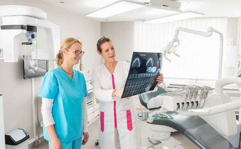
Imaging is a crucial tool in dentistry used to evaluate and record the size and shape of bone structures inside the mouth. Two-dimensional static imaging is commonly used, however, it does not allow dentists to properly gauge the depth of the bone structure. As a result, 3D imaging was developed during the early 1990’s. Since then, 3D X-ray imaging has been widely accepted as one of the best methods for evaluating dental problems and making an accurate diagnosis giving it a significant place in dentistry.
Why 3D Imaging is Necessary
With the availability of advanced technology, dental professionals today have the opportunity to enhance their treatment methods. Apart from consideration of cost and risk factors, the benefits of switching to 3D dental imaging are pretty obvious with both dentists and patients alike standing to gain from a better experience. With 3D X-ray imaging, practitioners can have a better view of dental anatomy from a wide range of angles with higher accuracy. It also helps the dentist to view the bone structures like adjacent root positions making it easier to find canals or root fractures.
Such imaging allows for a higher success rate for treatment as it provides a great deal of flexibility for dentists in terms of planning treatment, conducting root evaluations, and performing individual care. 3D x-ray imaging is both quick and accurate, enhancing reliability and ensuring a confident diagnosis. With 3D imaging, dentists receive an in-depth and complete assessment, allowing them to identify specific conditions and decide on the most efficient course of treatment. 3D dental imaging technology combined with advanced software help dentists prepare for definitive treatment plans that will best meet a patient’s individual needs.
Applications Of 3D Imaging
With 3D x-ray imaging, dentists are better able to:
- Determine normal or abnormal dental anatomy
- Identify location of nerves
- Detect dental problems
- Measure density and size of the jawbone
- View the position and orientation of teeth
- Detect impacted teeth
- More effectively plan surgical procedures
The imaging technology which has been in use since the early 2000’s in many parts of the world are also used for planning dental implants, viewing abnormal teeth, evaluating jaws and face, assessing cleft palate, diagnosing dental caries, cavities, root canals, and make a definitive diagnosis for any kind of dental trauma that has been sustained by a patient.
For more information on the benefits of 3D X-ray Imaging or to schedule an appointment, contact Foutz Family Dentistry today at 702-792-5929.
Dr. Barton H. Foutz, DDS
2510 Wigwam Parkway Suite 100 Henderson, NV 89074
(702) 792-5929